
When one thinks of X-rays and positron-emission tomography (PET) scans, their mind usually goes to broken bones and early cancer detection. Dr. Yuan-Chuan Tai, associate professor of radiology at Washington University in St. Louis (WUSTL), Missouri, estimates that more than 90 percent of PET scans are used for clinical oncological imaging. As a medical physicist and imaging scientist, his main focus with the technology is in clinical applications.
But building off several decades of research, Tai and scientists at the nearby Donald Danforth Plant Science Center in St. Louis are using PET scans and X-rays for another purpose: to study how plants behave.
Tracing plant mechanisms with PET scans
Tai has worked with PET scans for 26 years, but not always to advance the field of plant science. About 10 years ago, he says, the Department of Energy called for proposals from researchers who could use imaging technology to study how crops can produce energy. He applied for and received funding for plant-related imaging research. This was one opportunity that got him to do more work in that space, where he does a portion of his work still.
The Department of Energy has a wide interest in scientific research concerning “biology, environment, climate change [and] energy-producing technology,” including how plant mechanisms occur, Tai says. Much of this work has been made possible by plant imaging. For example: The Department of Energy was interested in how plants store carbon underground, and if there is a plant that can store massive amounts of carbon in its root system. All plants do this, Tai says, adding, “The goal is to understand the below-ground activity better to further improve the efficiency.”
With one of the largest PET research programs in the world, WUSTL uses four cyclotrons, or particle accelerators, to produce radioactive tracers, Tai says. These tracers allow PET to track their movement through plants, and scientists can look at processes such as carbon or nitrogen utilization, whether occurring under normal conditions or environmental changes. “If [scientists] need a new tracer to study a particular receptor, then we will work with chemists,” he says. “If they don't need a new receptor to study new a target — they just want to see where carbon goes — we don't need a chemist to be involved.”

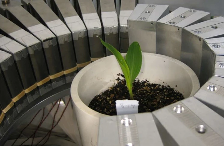
WUSTL also received funding from the National Science Foundation to develop scanners to image plants. “Most scanners had this horizontal bore where the patient lies down in the bed [to] slide in and out of the scanner,” Tai says. “Most plants grow vertically, so we built a device, and we put it inside a plant growth chamber. It has a vertical bore, so you can just drop a potted plant inside, and you can scan it from top to bottom, and we can control the environment, so we can study the plant.”
NSF funding also allowed Tai’s group to join the Plant Imaging Consortium (PIC) in 2014, which was formed by plant biologists and imaging specialists in Missouri and Arkansas. Tai was a co-principal investigator on the project until it ended in July. Work done through the PIC included PET scanning of tomato plants to study carbon movement through graft junctions, as well as how roots interact with fungus and bacteria in the soil.
Additionally, WUSTL works with the Danforth Center to study interactions between plants and fungi, Tai says. “There are certain strands of fungus that can help to convert phosphor in the soil into a form that plants can consume,” he says. “Fungi pick up the phosphor and convert it, then use it to exchange for the carbon with the plant. Plants get phosphor from fungus; fungus get carbon from plants. How they communicate and interact is an interesting scientific area.”
Mapping root structures using X-ray imaging
This specialized research is in full gear at Dr. Christopher Topp’s lab at the Danforth Center, says Keith E. Duncan, research scientist in the Topp Lab. “Chris’ lab has done nothing but study root systems since it's been around — could be alfalfa, could be sorghum, could be corn, could be soybean,” he says. “Any kind of root system — we're interested in how can we measure it, how can we evaluate it, without having to pull it up out of the soil."
The Danforth Center uses an X-ray tomography instrument that is encased in a lead box, weighing eight tons and measuring about nine square feet, Duncan says. Using nondestructive imaging and leaving the plant inside intact, the machine maps root structures — every aspect including thickness, deepness, number and complexity.
“They sell dozens of them to automotive plants and for electronics and aerospace to do nondestructive testing and imaging of very large or very dense, very complicated things,” Duncan says. “For us, we can take entire root systems that are growing — an entire corn plant or a soybean plant growing in a one or two or three-gallon pot — put the pot in there and scan the entire root system.”

Various scanning technologies offer several levels of resolution and provide different information when imaging plant root systems, Duncan says. PET scan technology can, in millimeter-resolution, take a full image of a corn or soybean root system in a one-gallon pot, although it needs to be separated into two separate top and bottom scans to cover the entire volume of the pot. Meanwhile, an X-ray tomography machine can image an entire root system in a single three-hour scan, while providing resolution in micrometers. It also shows scientists where plant roots are — something PET technology doesn’t do.
But the answer to many biological questions can’t be answered through the use of a single technology. “Working with Tai, and then working with computer scientists, we'll take that PET scan, overlay that with the X-ray scan, which shows you where all the roots are, and now, it's like having one map overlaid with another map,” Duncan says. “You have the map of the outline of the states, and then you overlay a transparency that has all the rivers.”
The Danforth Center also performs imaging with an X-ray microscope, which was the first device of its type to solely perform plant science in a plant science institute, Duncan says. It scans roots in soil, but at a much higher magnification than the larger X-ray tomography system, he says. In early August, Duncan used the X-ray microscope to identify and examine fungi inside root tissue — marking the first time anyone has ever done this using X-ray imaging.

Greenhouse applications
The Danforth Center paid for the X-ray microscope with financial help from Sumimoto Chemical and its wholly owned subsidiary Valent, the latter of which rents greenhouse space at the Danforth Center and works with it on research. In late July, Topp and Duncan brought virtual reality headsets to the grand opening of Valent BioSciences’ Biorational Research Center in Libertyville, Illinois, allowing attendees to virtually “stand inside” the root system of a plant. Tai also attended the event.
The Danforth Center performs blind experiments with growing media that includes products from Valent and its own wholly owned subsidiary, Mycorrhizal Applications, Duncan says. “We're [currently] working out the conditions whereby we can reliably image the root system and then overlay the PET information,” he says. “Once we have that down and can measure these things consistently, then we'll start comparing roots with and without the microbes, roots with and without the additives, roots with the additive plus and minus a drought condition or nitrogen starvation.”
Overlaying X-ray and PET scans is one process that makes data management and analysis so integral to the Topp Lab’s work, Duncan says. The lab employs as many computer scientists and mathematicians as it does biologists, with the former two groups working on projects like the virtual reality visualizations.
Duncan says he expects that Danforth Center research that looks at beneficial bacteria and fungi, and how they feed nutrients to plant roots, will have an impact on the ornamental industry. Although corn is a much different crop from ornamentals, the genetic controls for their root systems will often be similar. As the industry better understands root system architecture, it will be able to introduce useful soil additives.
Greenhouse growers, unlike field growers, have the ability to control their environment, Tai says. They could benefit from plant imaging research to add to that control. “If they have a better understanding of certain mechanisms underlying the growing cycle of plants, or to understand which species should be under certain kinds of growth conditions, then they can potentially fine-tune their greenhouse environment to maximize their yield,” he says. “I can imagine [this would help] them to have a better understanding of the greenhouse environment and how the conditions affect the productivity. That would actually have some commercial value.”

Explore the September 2018 Issue
Check out more from this issue and find your next story to read.
Latest from Greenhouse Management
- Don’t overlook the label
- Hurricane Helene: Florida agricultural production losses top $40M, UF economists estimate
- No shelter!
- Sensaphone releases weatherproof enclosures for WSG30 remote monitoring system, wireless sensors
- Profile Growing Solutions hires regional sales manager
- Cultural controls
- Terra Nova Nurseries shares companion plants for popular 2025 Colors of the Year
- University of Maryland graduate student receives 2024 Carville M. Akehurst Memorial Scholarship


